Marios Loukas from Grenada, WI and Albert van Schoor from Pretoria, RSA – two younger generation academic anatomists – have now been working with Jonathan Spratt, our consultant radiologist, to keep this atlas on the international cutting edge of anatomy as integrated into clinical medicine. This edition brings new coloured dissections, most of which were performed at the 3rd Hanno Boon Masterclass, held in 2016 at SGU, Grenada (see photo in the Acknowledgements). Over the last 40 years (8 edi-tions) the original book has moved with the times and ben-efited from the anatomical expertise of many international stars including Ralph Hutchings, Bari Logan and Professors John Pegington (UK), Sandy Marks (USA) and Hanno Boon (RSA) – all who made their own separate unique contribu-tions (see the sixth and seventh edition dedications and prefaces in the Student Consult eBook (For over 25 years Peter Abrahams has been the driving force keeping the first ever full-colour photographic dissection atlas so relevant today with updates on clinical practice and modern techniques, as well as the addition of numerous radiological modalities. A must have for any medical student.This new 8th edition based on the original McMinn Colour Atlas (1977) has now been updated and integrated with modern imaging anatomy, clinical case studies and 3D videos of most anatomical structures. It does not have enough MRI or CT images ,but that would haveĭefeated the original scope of this book as it is truly meant for medical
#F netter atlas of human anatomy pdf how to#
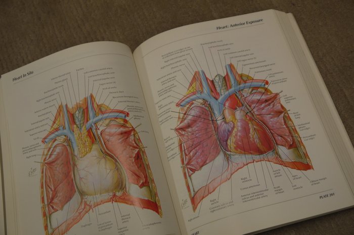
Thing now in this edition compared to 27 years ago is that it gives you anĮarly taste of radiology and how to identify different body parts from Relations, demonstrated by your Anatomy lecturer/demonstrator. Mostly now in medical schools youĪre given a dissected body revealing all the important structures and Most medical students will see in reality in there anatomy cathedrals asīodies are mostly preserved in formaldehyde and muscles,vessels,nerves The realistic pictures are perfect and something which ⭐ This book has never stoped surprising me,used it 27 years ago,now bought ⭐ This is an incredibly complete anatomy text. It can be related to shiping or paper quality instead of the book content: Reviews from Amazon users, collected at the time the book is getting published on UniedVRG.

Publications including Gray’s Anatomy Review, Gray’s Photographicĭissector, and Netter’s Introduction to Clinical Procedures.

Research and teaching and has published over 300 works in a variety of Served on the Student Evaluation and Promotion, and Faculty EvaluationĬommittees and was Chair of the Information Technology Committee at theĪmerican University of the Caribbean. Taught anatomy at the American University of the Caribbean and was Courseĭirector and Chairman of the Department of Anatomy for one year. School’s Department of Education and Development for seven years. Loukas taught anatomy, histology, and radiology at Harvard Medical Loukas is the University’s Dean of Research andĪssistant Dean of Basic and Allied Health Sciences. Guidance, the department publishes approximately 70 to 80 academic papers a Supervising research and inspiring scholarly activities. Responsible for managing the anatomy department, developing curriculum,

Georges University Medical School in 2008. Marios Loukas was appointed Chairman of the Department of Anatomical Abrahams and over 100 of his colleagues worldwide who have contributed to this unique collection of clinical anatomy images.ĭr. Learn from the culmination of over 45 years international clinical experience of Prof. – Master the 500 clinical conditions every physician should know by reviewing the associated clinical topics – featuring over 2000 additional clinical photos, radiological images, and case presentations not found in the printed book. – 200+ 3D scans,– allowing you to view the body in a more dynamic way to aid your understanding of dynamic anatomy.

This sets Abrahams’ and McMinn’s apart from any other atlases of human anatomy! Peter Abrahams and his team of leading international anatomists and radiologists link a vast collection of clinical images to help you master all the essential correlations between the basic science of anatomy and its clinical practice. Abrahams’ and McMinn’s Clinical Atlas of Human Anatomy, 8th Edition delivers the straightforward visual guidance you need to perform confidently in all examinations and understand spatial relationships required during your medical training, while also acquiring the practical anatomical knowledge needed for your future clinical career.


 0 kommentar(er)
